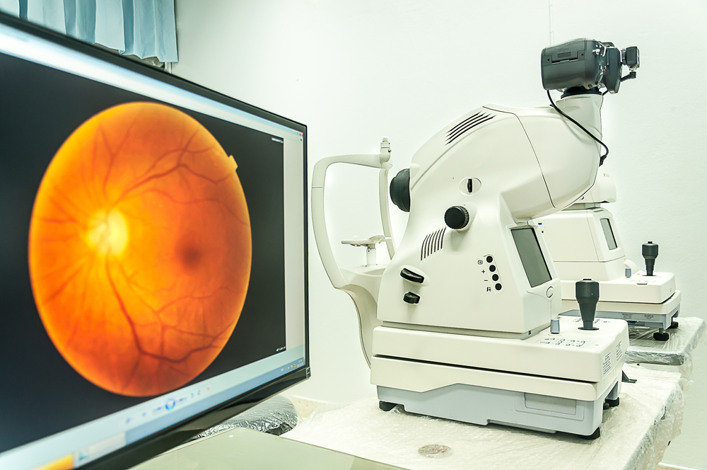
A comprehensive eye exam involves several tests, including fundus photography. Eye doctors perform this test to catch eye conditions early before they harm a patient’s health.
Uses of a Fundus Camera
Your ophthalmologist may order retinal imaging to detect, assess, and treat various eye disorders, such as:
Glaucoma
Diabetic retinopathy
Hypersensitive retinopathy
Age-related macular degeneration
Optic atrophy
Congenital rubella
Retinoblastoma
Congenital glaucoma
Color vision deficiencies
Cancer of the eye
Papilledema
Toxoplasmosis
Congenital eye defect
Fundus Camera
A fundus camera, also known as a retinal camera, is a low-power microscope with a camera designed to photograph the eye’s interior surface. That includes the retina, optic disc, retinal vasculature, macula, and fundus.
Many digital retinal cameras have features like angiography and red-free imaging, color, angle variations, and LCD monitors. The images captured by the camera allow for detailed image study and quick transfer, as well as side-by-side comparison. They are essential in the accurate identification and treatment of various eye diseases.
What Is Fundus Photography?
Retinal photography is a painless and quick way to look inside the eye and track ocular and vision changes. A fundus camera is a noninvasive diagnostic tool designed to produce high-resolution, colored images of the retina, blood vessels, and optic nerve.
Retinal imaging is a vitally important test, but it is not a substitute for regular comprehensive eye exams. Rather, it allows a more expansive and precise view of the retina for early detection of eye conditions.
Do You Need a Retinal Imaging Test?
Eye doctors commonly perform fundus photography as part of their regular eye exams. Your eye doctor may recommend this test if you experience vision changes to rule out eye conditions that may be detrimental to your retina. Additionally, you may need to undergo this test if you have any of the following conditions:
Macular degeneration
Diabetes
Hypertension
High cholesterol
Retinal toxicity
Glaucoma
The only visible blood vessels in the body are the retinal blood vessels. That is why doctors use digital fundus photography to detect general health conditions.
What to Expect During a Digital Retinal Imaging Test
Before taking digital photographs of your retina using a fundus camera, your eye doctor may dilate your pupils using special eye drops. You will then need to place your forehead and chin on supportive rests to help keep your head still.
Next, your eye doctor will ask you to open your eyes as wide as possible as you stare at an object ahead of you. You will see a bright flash when the doctor takes the photograph to capture high-definition images of the inner surface of your eye. The photos will appear on a computer screen for your doctor to review.
What to Expect After the Test
Often, retinal imaging tests do not require the application of eye drops. However, it would be best to rest for a few minutes for the effects of the flash to wear off. The noninvasive test takes only a couple of minutes.
For more on fundus photography, call Primary Vision Care today at one of our 5 locations in Ohio: Lancaster 740.654.9909, Newark, 740.299.1155, Mount Vernon 740.393.6010, Wilmington 937.382-4933, & Waynesville 513.897.2211.


